Conduction system of the heart

Conduction system of the heart is a complex of anatomical formations of the heart (nodes, bundles and fibers) consisting of atypical muscle fibers (cardiac conduction muscle fibers). It provides coordinated work of different parts of the heart (atria and ventricles) aimed at providing normal cardiac activity.
Conduction system of the heart consists of two interrelated parts: sinoatrial and atrioventricular.
The sinoatrial part includes sinus node, three fast-conducting bundles, linking the sinus node to the atrioventricular node, and the intraatrial fast-conducting bundle that connects the sinus node to the left atrium.
The atrioventricular part consists of the atrioventricular node, the bundle of His (includes the common trunk and three branches: the left anterior, the left posterior and right) and the Purkinje fibers.
Conduction system of the heart begins with a sinus node, which is located subepicardially in the upper part of the right atrium between the mouthes of the cava veins. It is a bundle of specific tissues. Its length is 10-20 mm, thickness is 3-5 mm. The node consists of two types of cells: P-cells (they generate excitation impulses), T-cells (they conduct impulses from the sinus node to the atria).
The atrioventricular node is located in the lower part of the right atrium to the right of the interatrial septum, next to the mouth of coronary sinus. Its length is 5 mm, thickness is 2 mm. By analogy with the sinus node, the atrioventricular node also consists of P-cells and T-cells.
The atrioventricular node passes into the bundle of His, which consists of the penetrating (initial) and branching segments. The initial part of the bundle of His has no contacts with the contractile myocardium and is not sensitive to the lesion of the coronary arteries, but it is easily involved in the pathological processes occurring in the fibrous tissue that surrounds bundle of His. The length of the bundle of His is 20 mm.
The bundle is divided into 2 branches (right and left). Further, the left branch of the bundle of His is divided into two hemifascicles. The result is the right branch and two hemifascicles of the left branch, which descend down both sides of the interventricular septum. The right branch goes to the right ventricle of the heart. As for the left branch, the opinions of the researchers here diverge. It is believed that the anterior hemifascicle of the left branch of the bundle of His provides fibers to the anterior and lateral walls of the left ventricle; the posterior hemifascicle provides fibers to the posterior wall of the left ventricle and the lower sections of the lateral wall.
The intraventricular conduction system can be considered as a system consisting of 5 main parts: the bundle of His, the right bundle branch, the main branch of the left bundle branch, the anterior hemifascicle of the left bundle branch, the posterior hemifascicle of the left bundle branch. The right bundle branch and the anterior hemifascicle of the left bundle branch are the thinnest, therefore vulnerable. Further, in terms of vulnerability, there are the main branch of the left bundle branch, the bundle of His, the posterior hemifascicle of the left bundle branch.
The branches of the intraventricular conduction system gradually splits out to smaller branches and gradually pass into the Purkinje fibers, which bind directly to the contractile myocardium of the ventricles, permeating the entire muscle of the heart.
The contractions of the cardiac muscle (myocardium) occur due to the impulses arising in the sinus node and spreading through the conduction system of the heart: through the atria, the atrioventricular node, the bundle of His, Purkinje fibers. Impulses are conducted to the contractile myocardium.
In the heart there are many cells that have the function of automatism:
- Sinus node (first-order automatic center) has the greatest automatism;
- Atrioventricular node (automatic center of the second order);
- Bundle of His and its branches (automatic center of the third order).
Normally, there is only one pacemaker. It's a sinus node, the impulses from which spread to the underlying sources of automatism before they complete the preparation of the next excitation impulse, and destroy this process of preparation. Simply put, the sinus node is normally the main source of excitation, suppressing similar signals in automatic centers of the second and third order.
Automatic centers of the second and third order manifest their function only in pathological conditions, when the automatism of the sinus node decreases, or their automatism increases.
The automatic center of the third order becomes the pacemaker at decrease of functions of the automatic centers of the first and second orders, and also at increase of own automatic function.
The conduction system of the heart can conduct impulses not only in the forward direction, from the atria to the ventricles (antegrade), but also in the opposite direction, from the ventricles to the atria (retrograde).
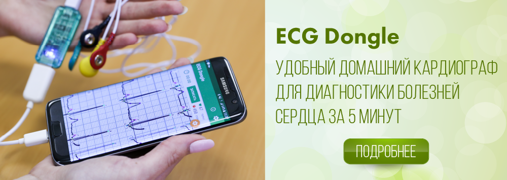
It is possible to identify violations of the cardiac conduction system at home using the ECG Dongle [1].